#Galeazzi fracture diagram how to
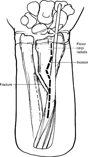
Plate and screw fixation is the preferred method, and the review describes the technique in detail, including how to address comminution. Treatment of AdultsĪdults with a Galeazzi fracture require open surgery for anatomic and rigid fixation of the radius shaft and stabilization of the DRUJ. Computed tomography (CT) or a radiograph of the contralateral wrist can be useful for assessing the DRUJ. In some cases, injury to the DRUJ is purely ligamentous. More than 5 mm of shortening of the radius relative to the ulna.

Dislocation of the radius relative to the ulna (on a true lateral view).Widening of the DRUJ space (on a true AP view).Fracture at the base of the ulnar styloid.Neurovascular damage is rare.Įssential imaging includes good anteroposterior (AP) and lateral radiographs of the forearm, as well as orthogonal views of the wrist and elbow. Galeazzi patients usually present with swelling and deformity of the forearm. Mudgal, MD, in the Hand and Arm Center at Massachusetts General Hospital, reviewed the evaluation and treatment of Galeazzi fractures, which are sometimes called Piedmont fractures, reverse Monteggia fractures or Darrach–Hughston–Milch fractures. In Hand Clinics, orthopedic surgeons Rohit Garg, MD, MBBS, and Chaitanya S. These injuries usually occur as a result of high-energy trauma (e.g., motor vehicle accident, sports injury or a fall from a height) or a fall on an outstretched and pronated hand. J Hand Surg Am.Error: Please enter a valid email address.įractures of the radius shaft associated with a dislocation of the distal radioulnar joint (DRUJ) are called Galeazzi fractures. A Historical Report on Riccardo Galeazzi and the Management of Galeazzi Fractures. William Darrach, MD: his life and his contribution to hand surgery. Journal of Bone and Joint Surgery (British). (Of a particular traumatic syndrome of the forearm’s skeleton). Atti e memorie della Società lombarda di chirurgia. Di una particolare sindrome traumatica dello scheletro dell’ avambraccio. Archiv fur orthopadische und Unfall Chirurgie 1934 35:557–562. Uber ein besonderes Syndrom bei Verletzungen im Bereich der Unterarmknochen. Fracture of the lower end of the radius associated with fracture or dislocation of the lower end of the ulna. Forward dislocation at the inferior radio-ulnar joint with fractures of the lower third of the radius. In: A treatise on dislocations, and on fractures of the joints. Compound dislocation of the ulna with fracture of the radius. Archiv fur orthopadische und Unfall Chirurgie 1934

He reduced the radial shaft fracture by pulling on the thumb with the forearm in the supinated position, and the ulnar head by radially deviating the wrist, and maintained the reduction using a plaster of Paris cast. ġ925 – Further attempts at open reduction by Wilson and Cochrane (1925) and Milch (1926) with triangular fibrocartilage repair with a fascial graft – with poor outcomesġ934 – Riccardo Galeazzi described radial shaft fracture with associated DRUJ dislocation and published his experience of 18 cases. Reviewed 18 months later, found patient unable to pronate/supinate – recommended subperiosteal excision of the distal end of the ulna for delayed cases of ulnar head dislocation (Darrach procedure) ġ922 – Homans and Smith attempted open reduction of the radius fracture and applied a splint, but found a tendency for angulation and delayed union in cases associated with a dislocation of the radioulnar joint. He attempted closed reduction unsuccessfully prior to open reduction and splinting in pronation. In his Treatise on dislocations, and on fractures of the joints Astley Cooper description 1822ġ912 – William Darrach (1876–1948) reported a 20 year old male presenting with an 8 week old injury with ‘ fracture of the radial shaft 2.5 inches above the lower margin with anterior bowing and a forward dislocation of the head of ulna‘. Copper describes cases of radius fracture with DRUJ disruption and ulna dislocation Compound dislocation of the ulna with fracture of the radius. 1822 – Fracture pattern first described by Sir Astley Cooper some 110 years prior to Galeazzi’s publication.


 0 kommentar(er)
